Raman Microscopy
Raman Microscopy
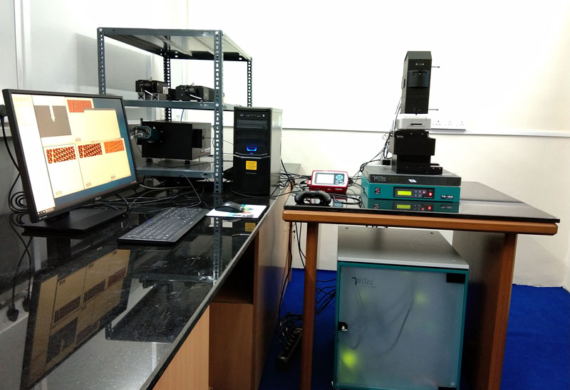
Confocal Raman Microscope with AFM imaging (WiTec alpha 300, Germany) installed at PSG Institute of Advanced Studies is an integrated system with high resolution nanoimaging capability. It can be mainly used for chemical analysis at sub-micrometer scale.
The confocality of the optical design facilitates excellent depth resolution and makes the generation of 3D Raman images and depth profiles easily with a Raman Spectral range of 50 cm-1 – 4000 cm-1. Confocal micro-Raman spectroscope has features such as, Single-point spectra acquisition, Single-point depth profiling and 3D imaging and depth profiling. The advantage of an integrated system with a combination of ultra-high topographic and lateral resolution of AFM with Raman spectroscopy provides a more comprehensive understanding of the samples; not only the chemical information, but also structural and topographic information acquired at the same time and from the same sample area.
The system is integrated with a motorized stage as well as high resolution piezo XYZ mapping Stage with AFM imaging capability. The instrument is equipped with 355 nm and 532 nm lasers as UV Excitation Laser source for PL analysis and VIS Excitation Laser source for Raman analysis respectively. Using optical pre-inspection by means of various illumination and detection techniques (e.g. bright field, dark field, polarization, fluorescence, etc.) the area of interest for the AFM/Raman imaging can be easily determined. The system is capable of measuring the Raman spectra of wide variety of samples including amorphous, crystalline, polymer or liquid/biological samples.
Contact
- Dr. P. Biji
- Mail : pbm@psgias.ac.in
Who Can Use This Facility?
- University researchers
- Industrial R&D teams
- Independent scientists
- Startups and material science innovators
Booking & Usage
Operating Hours: Monday to Friday, 9:00 AM – 5:00 PM
Booking: Prior appointment required
Charges: Nominal analysis fee applicable (Download latest [fee structure PDF])